Medical image analysis

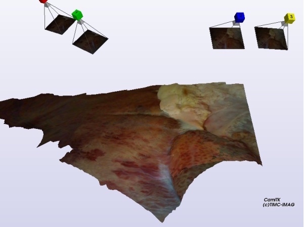
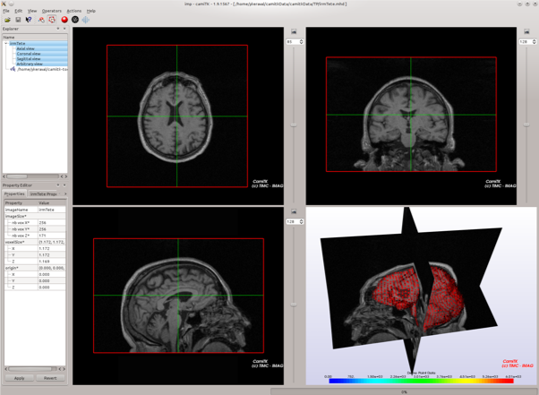
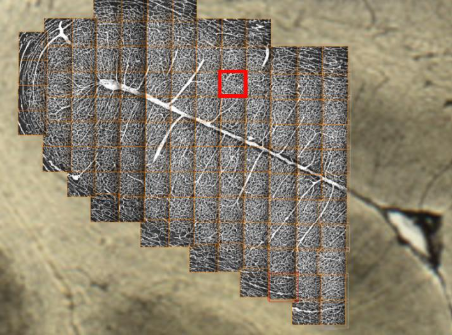
Medical image analysis is the common thread running through my entire career — long before the term “AI” was superimposed on it. From confocal microscopy of the cerebral cortex to intraoperative ultrasound, from CT imaging in prostate brachytherapy to cardiac MRI, each project has asked the same fundamental question: how do you extract useful, robust information from an image that a clinician can actually use, in a real clinical context?
This theme brings together methodological contributions — chamfer distances, skeletonisation, registration, segmentation — and concrete clinical applications in orthopaedics, oncology and cardiology, spanning from my PhD thesis to the most recent projects.
Projects & publications
Publications
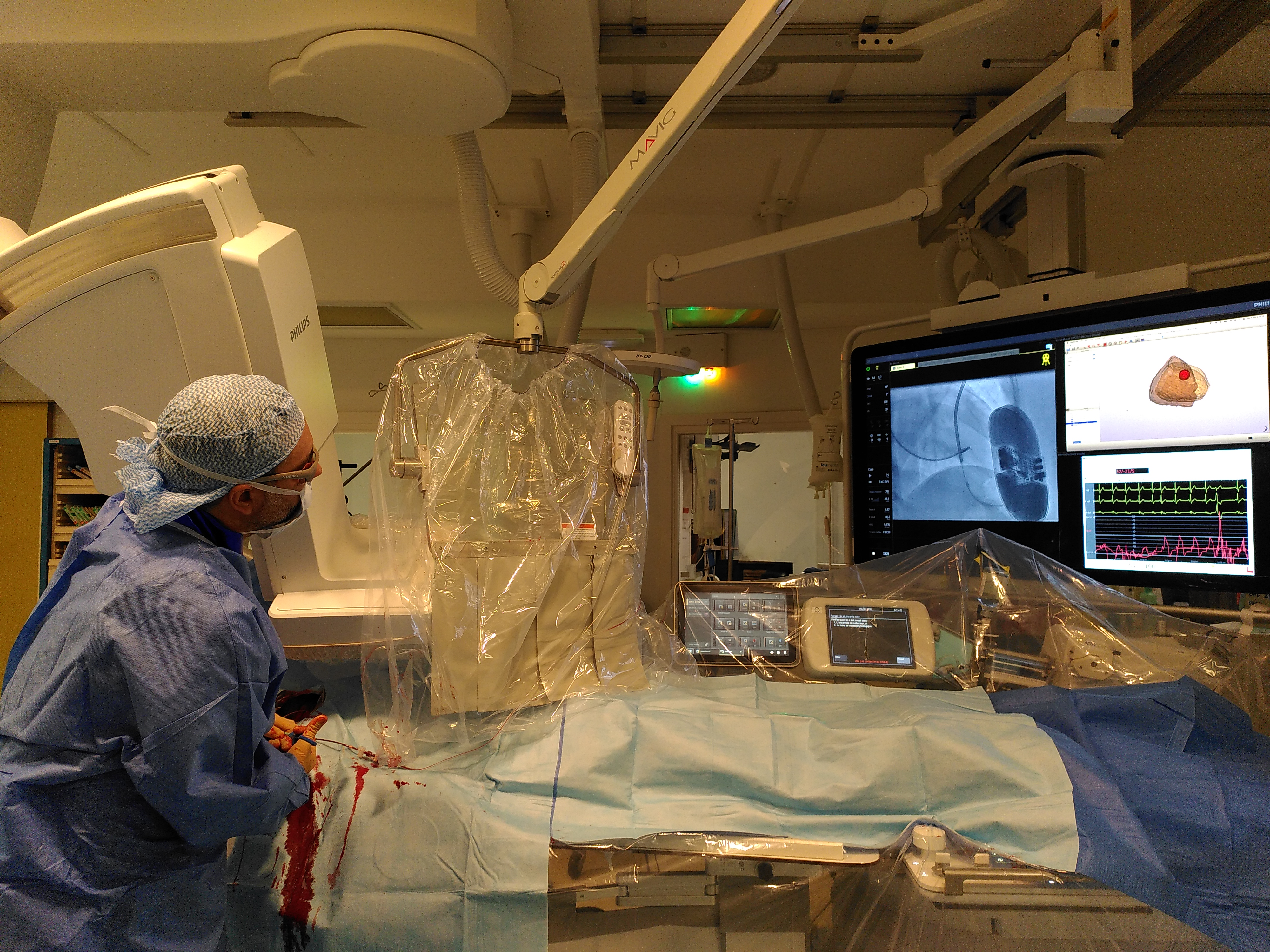
- 2024 Barone-Rochette Gilles, MD,,Erwan Lecesne, MSc,,Antoine Simon, PhDet al. —New Method CMR-Guided Endomyocardial Biopsy in Suspicion Context of Isolated Cardiac Sarcoidosis · *Circulation: Cardiovascular Imaging, vol 17, no 4*
- 2023 Erwan Lecesne,Antoine Simon,Mireille Garreauet al. —Segmentation of cardiac infarction in delayed-enhancement MRI using probability map and transformers-based neural networks · *Computer Methods and Programs in Biomedicine, vol 242*
- 2023 Lecesne Erwan,Simon Antoine,Garreau Mireilleet al. —Transformers-Based Neural Network for Cardiac Infarction Segmentation in Delayed-Enhancement MRI · *2023 IEEE Twelfth International Conference on Image Processing Theory, Tools and Applications (IPTA)*
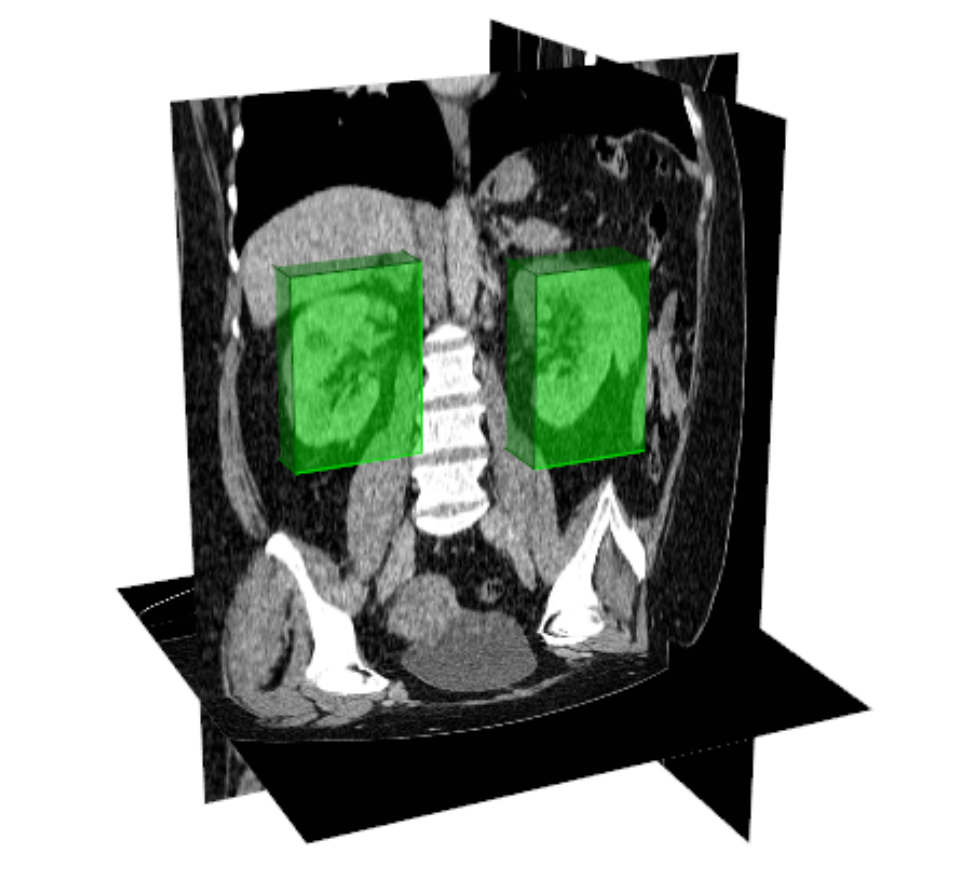
- 2017 Samarakoon Prasad N,Promayon Emmanuel,Fouard Céline —Light Random Regression Forests for automatic multi-organ localization in CT images · *2017 IEEE 14th International Symposium on Biomedical Imaging (ISBI 2017)*
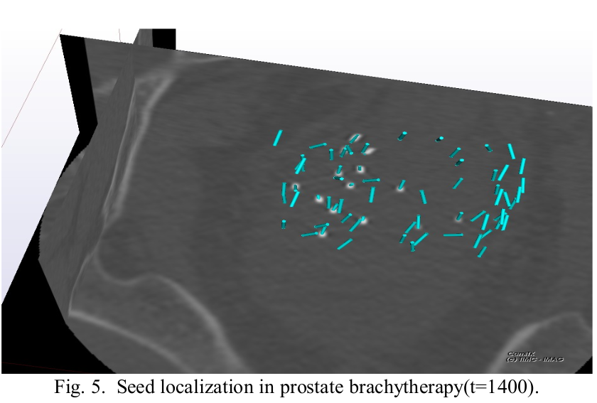
- 2015 Nguyen Huu-Giao,Fouard Céline,Troccaz Jocelyne —Segmentation, separation and pose estimation of prostate brachytherapy seeds in CT images · *IEEE Transactions on Biomedical Engineering, vol 62, Issue 8*
- 2014 Meneu François,Nguyen Huu-Giao,Fouard Célineet al. —Impact de l’orientation des grains iode 125 dans l’évaluation de la dosimétrie à 1 mois d’une curiethérapie prostatique · *Cancer/Radiothérapie, vol 18, Issue 5-6*
- 2014 Nguyen Huu-Giao,Fouard Céline,Meneu Françoiset al. —Automatic 3D seed location and orientation detection in CT image for prostate brachytherapy · *Biomedical Imaging (ISBI), 2014 IEEE 11th International Symposium on*
- 2014 Saramakoon Prasad,Promayon Emmanuel,Fouard Céline —Fully Automatic Organ Localization in Medical Images Using Improved Random Regression Forests · *Proceedings of Surgetica 2014*
- 2013 Meneu François,Nguyen Huu-Giao,Chartier Pascalet al. —Consideration of seeds orientation in prostate brachytherapy and dosimetric analysis · *Physica Medica Vol 29*
- 2010 Schers Jonathan,Fouard Céline,Troccaz Jocelyne —Non invasive ultrasound-based bone tracking · *Computer Assisted Orthopaedic Surgery (CAOS 2010)*
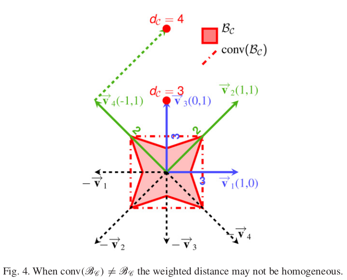
- 2007 Fouard Céline,Strand Robin,Borgefors Gunilla —Weighted distance transforms generalized to modules and their computation on point lattices · *Pattern Recognition Vol 40 Issue 9*
- 2006 Fouard Céline,Gedda Magnus —An Objective Comparison between Gray Weighted Distance Transforms and Weighted Distance Transforms On Curved Spaces · *Proceedings of DGCI 2006*
- 2006 Strand Robin,Nagy Benedek,Fouard Célineet al. —Generating Distance Maps with Neighbourhood Sequences · *Discrete Geometry for Computer Imagery*
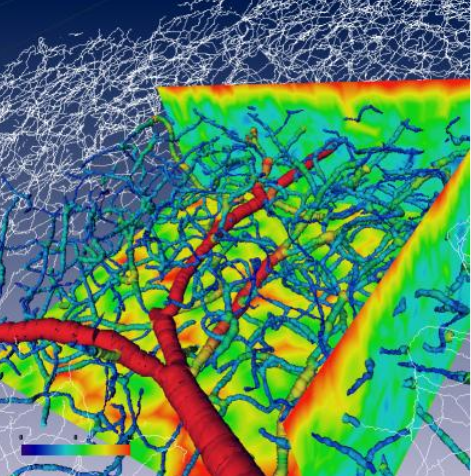
- 2006 Fouard Céline,Malandain Grégoire,Prohaska Steffenet al. —Blockwise processing applied to brain microvascular network study · *IEEE Transactions on Medical Imaging ( Volume: 25, Issue: 10, October 2006) *
- 2006 Cassot Francis,Lauwers Frederic,Fouard celineProhaska Steffenet al. —A Novel Three-Dimensional Computer-Assisted Method for a Quantitative Study of Microvascular Networks of the Human Cerebral Cortex · *Microcirculation*
- 2005 Fouard Céline,Malandain Grégoire —3-D chamfer distances and norms in anisotropic grids · *Image and Vision Computing*
- 2005 Fouard Céline —Extraction de paramètres morphométriques pour l'étude du réseau micro-vasculaire cérébral · Thèse de doctorat, *Université de Nice Sophia Antipolis*
- 2003 Fouard Céline,Malandain Grégoire —Systematized calculation of optimal coefficients of 3-D chamfer norms · *Discrete Geometry for Computer Imagery, 11th International Conference, DGCI 2003*